More About Ct Scan Melbourne
Table of ContentsThe Buzz on Cat Scan MelbourneThe 8-Minute Rule for Ct Scan MelbourneThe Best Strategy To Use For X-ray Melbourne
Dr. Macintyre's X-Ray Movie (1896) Radiology is the clinical self-control that utilizes medical imaging to identify as well as treat diseases within the bodies of pets, consisting of people. A selection of imaging techniques such as X-ray radiography, ultrasound, computed tomography (CT), nuclear medication consisting of positron emission tomography (PET), as well as magnetic vibration imaging (MRI) are utilized to identify or deal with illness.The contemporary method of radiology involves numerous different healthcare occupations working as a team. The radiologist is a medical doctor who has actually completed the proper post-graduate training as well as analyzes clinical pictures, interacts these searchings for to various other physicians using a report or verbally, and makes use of imaging to carry out minimally intrusive clinical procedures.

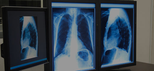
The X-rays are projected through the body onto a detector; a picture is developed based upon which rays go through (as well as are identified) versus those that are soaked up or scattered in the individual (and also therefore are not spotted). Rntgen uncovered X-rays on November 8, 1895 as well as received the initial Nobel Prize in Physics for their discovery in 1901.
The X-rays that go through the person are filtered with a device called an grid or X-ray filter, to minimize scatter, and strike an undeveloped film, which is held snugly to a screen of light-emitting phosphors in a light-tight cassette. The film is after that developed chemically as well as a photo shows up on the film.
In the two newest systems, the X-rays strike sensors that converts the signals produced into digital details, which is transmitted and exchanged a picture displayed on a computer system display. In digital radiography the sensing units form a plate, however in the EOS system, which is a slot-scanning system, a linear sensing unit up and down scans the client.
An Unbiased View of X-ray Melbourne
Because of its accessibility, rate, as well as lower expenses compared to other modalities, radiography is typically the first-line test of option in radiologic diagnosis. Likewise in spite of the huge quantity of data in CT scans, MR scans and other digital-based imaging, there are several disease entities in which the timeless medical diagnosis is obtained by plain radiographs - Ct Scan Melbourne.
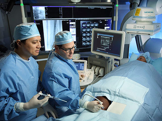
Mammography and DXA are 2 applications of low energy projectional radiography, made use of for the analysis for breast cancer cells and osteoporosis, respectively. Fluoroscopy and also angiography are special applications of X-ray imaging, in which a fluorescent display and image intensifier tube is attached to a closed-circuit tv system.:26 This allows real-time imaging of frameworks in motion or increased with a radiocontrast representative.
Two radiocontrast representatives are currently in common usage. Barium sulfate (BaSO4) is offered orally or rectally for evaluation of the GI system. Iodine, in multiple proprietary kinds, is given by oral, rectal, vaginal, intra-arterial or intravenous courses. These radiocontrast representatives strongly soak up or spread X-rays, and also together with the real-time imaging, enable demonstration of dynamic procedures, such as peristalsis in the digestion tract or blood flow in arteries as well as blood vessels.
With fast administration of intravenous comparison throughout the CT scan, these great information pictures can be rebuilded into three-dimensional (3D) pictures of carotid, cerebral, coronary or other arteries. The intro of computed tomography in the early 1970s revolutionized analysis radiology by offering Medical professionals with photos of actual three-dimensional anatomic frameworks.
8 Easy Facts About Cat Scan Melbourne Explained
The first ultrasound pictures were static as well as two-dimensional (2D), however with contemporary ultrasonography, this contact form 3D restorations can be observed in genuine time, effectively coming to be "4D". Since ultrasound imaging methods do not employ ionizing radiation to generate photos (unlike radiography, as well as CT scans), they are usually taken into consideration much safer and also are consequently more typical in obstetrical imaging.
Ultrasounds serves as a guide to doing biopsies to minimise damages to surrounding tissues as well as in you can check here water drainages such as thoracentesis. Small, portable ultrasound gadgets currently replace peritoneal lavage in injury wards by non-invasively analyzing for the existence of Our site inner blood loss and also any interior organ damages. Substantial interior blood loss or injury to the significant organs may need surgical procedure and also fixing. Ct Scan Melbourne.